
Rétablissement hypothétique de la vie de MOR 7029. Notez que la maladie pulmonaire infectant cet animal n’aurait pas été extérieurement évidente, mais les symptômes extérieurs probables de type pneumonie auraient inclus, entre autres, la toux, la respiration laborieuse, l’écoulement nasal, la fièvre et la perte de poids. Crédit : Woodruff, et al. (2022) et Corbin Rainbolt.
La découverte est la première occurrence d’une infection respiratoire de type aviaire chez un dinosaure non aviaire.
Les restes fossilisés d’un diplodocide immature – un grand dinosaure sauropode herbivore à long cou, comme… “.Brontosaurus” – pourraient fournir la première preuve d’une infection respiratoire unique chez un dinosaure, selon une étude publiée dans la revue Scientific Reports. Ces résultats améliorent notre compréhension des maladies qui affectaient les dinosaures.
Le spécimen, surnommé “Dolly”, a été découvert dans le sud-ouest du Montana, aux États-Unis, et date de la fin du Jurassic Period of the Mesozoic Era (approximately 150 million years ago). Cary Woodruff of the Great Plains Dinosaur Museum in Malta, along with his colleagues, examined three of the cervical vertebrae (the bones from the neck) from Dolly and identified never-before-seen abnormal bony protrusions that had an unusual shape and texture. These protrusions were located in an area of each bone where they would have been penetrated by air-filled sacs. These air sacs would have ultimately connected to Dolly’s lungs and formed part of the dinosaur’s complex respiratory system. CT imaging of the irregular protrusions revealed that they were made of abnormal bone that most likely formed in response to an infection.
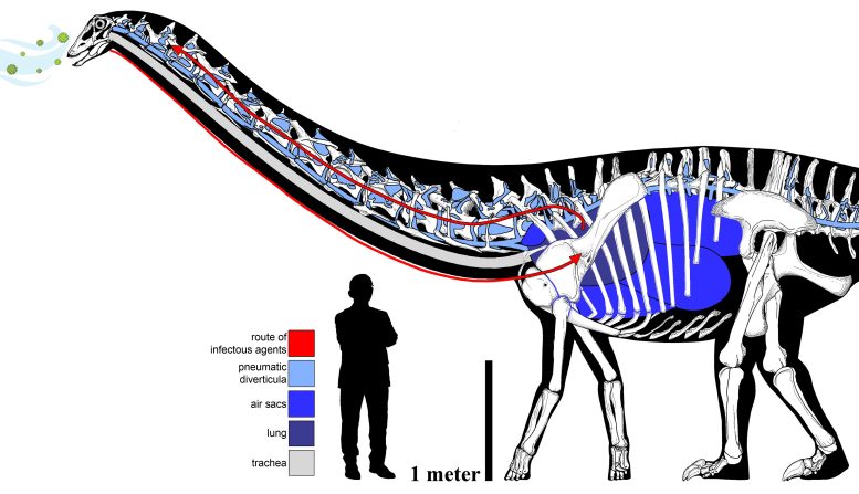
The elaborate and circuitous pulmonary complex of the sauropod, with the hypothetical route of infectious pathway in MOR 7029. Human scale bar is the profile of a man standing 170cm tall. Credit: Woodruff, et al., and Francisco Bruñén Alfaro
“Given the likely symptoms this animal suffered from, holding these infected bones in your hands, you can’t help but feel sorry for Dolly,” Woodruff said. “We’ve all experienced these same symptoms – coughing, trouble breathing, a fever, etc. – and here’s a 150-million-year-old dinosaur that likely felt as miserable as we all do when we’re sick.”
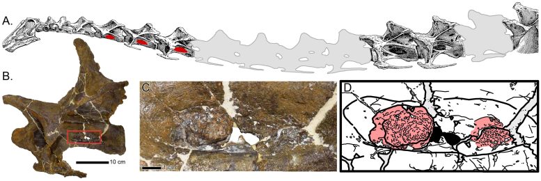
Abnormal bony growth in MOR 7029. (A) Schematic map of the neck of Diplodocus, with the abnormal bone growth denoted in red. (B) Neck vertebra of MOR 7029 with a red box highlighting the abnormal structure; close up in (C) with interpretative drawing in (D) (abnormal structure in red). Credit: Woodruff, et al.
Based on the location of these abnormal bony protrusions, the researchers suggest that they formed in response to a respiratory infection in Dolly, which ultimately spread into these neck vertebrae via the air sacs and caused the irregular bone growths. The authors speculate that this respiratory infection could have been caused by a fungal infection similar to aspergillosis, a common respiratory illness that affects birds and reptiles today and can lead to bone infections. In addition to documenting the first occurrence of such a respiratory infection in a dinosaur, this fossilized infection also has important anatomical implications for the respiratory system of sauropod dinosaurs.
“This fossil infection in Dolly not only helps us trace the evolutionary history of respiratory-related diseases back in time, but gives us a better understanding of what kinds of diseases dinosaurs were susceptible to,” Woodruff said.
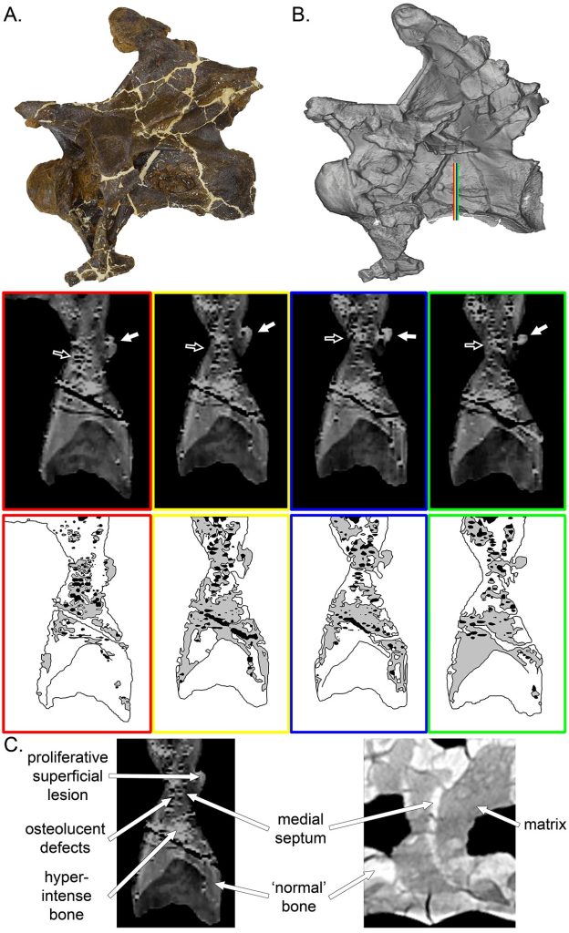
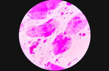
CT scans of infected vertebra from Dolly. Photograph and scan model of the infected vertebra (A & B respectively). The colored lines in (B) correspond to the scan slices (and scan interpretative drawings below). White arrows point to the externally visibly abnormal bone growth, while black arrows denote the internal irregularities. (C) Comparison of the abnormal tissue composition of Dolly (left), compared to that of a ‘normal’ sauropod (right). Credit: Woodruff, et al.
The researchers suggest that if Dolly had been infected with an aspergillosis-like respiratory infection, it likely experienced flu or pneumonia-like symptoms such as weight loss, coughing, fever, and breathing difficulties. As aspergillosis can be fatal in birds if untreated, a potentially similar infection in Dolly could have ultimately caused the death of the animal, they add.
In addition to Woodruff, the research team included a paleopathologist/veterinarian, Ewan Wolff (University of New Mexico); a veterinarian, Sophie Dennison (TeleVet Imaging Solutions, Oakton, Va.); and two paleontologists who are also medical anatomists, Mathew Wedel (Western University of Health Sciences, Pomona, Calif.) and Lawrence Witmer (Ohio University Heritage College of Osteopathic Medicine, Athens, Ohio).
Reference: “The first occurrence of an avian-style respiratory infection in a non-avian dinosaur” by D. Cary Woodruff, Ewan D. S. Wolff, Mathew J. Wedel, Sophie Dennison and Lawrence M. Witmer, 10 February 2022, Scientific Reports.
DOI: 10.1038/s41598-022-05761-3